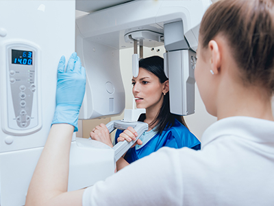
The image shows a person receiving a medical procedure, possibly a dermatological treatment, under the supervision of a healthcare professional using a magnifying device, set against a blue background with a medical table and equipment visible in the foreground.

Digital Radiography

Understanding Digital Radiography: A Modern Approach to Dental Imaging

Digital radiography replaces traditional film with electronic sensors and computer processing to capture images of your teeth, jaws, and surrounding tissues. Instead of waiting for film to be developed, the sensor transmits the image directly to a computer where it appears almost instantly. This shift from analog to digital makes routine x-rays quicker, more flexible, and easier for clinicians and patients to review together.

At its core, digital radiography uses the same basic principles as conventional x-rays—controlled x-ray exposure produces an image—while taking advantage of modern digital capture and display methods. The equipment includes small intraoral sensors or external digital panels for broader views, plus software that stores, enhances, and organizes images as part of a patient’s electronic health record. Because images are digital files, they can be manipulated for clarity and saved without taking up physical storage space.

For patients, the benefits of this technology are easy to see: faster appointments, clearer explanations from your dental team, and a reduced environmental footprint compared with chemical-based film processing. For clinicians, digital imaging is a diagnostic tool that integrates smoothly with other digital systems in the office, helping the team make timely, well-informed treatment decisions.

Minimizing Exposure: How Digital Sensors Support Safer Imaging

One of the most important advantages of digital radiography is its ability to produce high-quality images with less radiation than traditional film techniques. Modern sensors are more sensitive to x-rays, which means the same diagnostic information can often be obtained with a lower dose. Reducing exposure supports a cautious, patient-centered approach to imaging while still delivering the detail clinicians need.

Safety in dental imaging also depends on technique and equipment calibration. Dental teams follow professional guidelines to determine when an x-ray is necessary and which views are most appropriate, applying the ALARA principle—keeping radiation "as low as reasonably achievable"—to every decision. When combined with properly maintained digital equipment, these practices contribute to safer care for patients of all ages.

Beyond dose reduction, digital systems often include features that aid safe use: automatic exposure controls, software alerts for correct positioning, and image review capabilities that reduce the need for repeat exposures. These safeguards help ensure that each image is acquired efficiently and effectively the first time.

Sharper Detail, Smarter Diagnosis: Clinical Advantages of Digital Images

Digital images can be adjusted instantly to highlight specific details—brightness, contrast, magnification, and measurement tools allow clinicians to examine structures more closely than a static film might permit. Enhanced visualization is especially helpful for spotting early decay, assessing root structures, evaluating bone levels around teeth, and planning restorative or surgical procedures with greater confidence.

Because images are available immediately, clinicians can discuss findings with patients during the same visit. This real-time review makes it easier to explain treatment needs, show progress over time, and involve patients in decisions about their oral health. Digital tools also support side-by-side comparisons of past and present images, enabling clear tracking of changes and outcomes.

Digital radiography supports a broad range of clinical workflows, from routine bitewings and periapical images to panoramic or cone-beam views when more comprehensive imaging is required. The adaptability of digital systems helps the dental team select the right view for each diagnostic question while maintaining consistent image quality across different types of exams.

Records, Security, and Collaboration: The Administrative Strengths of Going Digital

Storing images as digital files simplifies recordkeeping and improves access. Instead of filing and retrieving physical films, images become part of the electronic chart and can be retrieved instantly during a visit. This convenience reduces administrative overhead and helps the office maintain a complete, well-organized clinical record for each patient.

Digital files are also easier to share securely with other clinicians when a second opinion or specialist consultation is needed. Secure transfer methods preserve image fidelity and patient confidentiality, allowing collaborators to review the same high-resolution files and provide coordinated recommendations. This interoperability supports more cohesive care when multiple providers are involved.

From a practice standpoint, digital systems often include backup and audit features that protect records against loss and help practices comply with privacy and data-security expectations. These safeguards ensure that important diagnostic information remains available and intact over time, supporting continuity of care across appointments and phases of treatment.

What Patients Can Expect During a Digital X-Ray Appointment

A digital x-ray appointment is fast and straightforward. For intraoral images, a small sensor is positioned inside the mouth and held briefly while the image is taken. Because the sensor is thinner and more flexible than traditional film, many patients find it more comfortable. Panoramic or extraoral digital panels are noninvasive and require only brief, simple positioning.

After the exposure, the image appears on the clinician’s monitor within seconds. Your dental team will use on-screen tools to adjust the image for clarity and point out any areas of interest. This immediate feedback helps you understand what the clinician is seeing and lets the team answer questions and outline next steps during the same appointment.

If you have concerns about imaging—related to pregnancy, prior exposure, or medical history—be sure to discuss them with your dental team before the x-ray. Clinicians will review your history, apply appropriate precautions, and choose the imaging approach that balances diagnostic benefit with patient comfort and safety.

Digital radiography has become a cornerstone of modern dental care, offering faster results, clearer images, and a safer, more efficient experience for patients and clinicians alike. At Dental Illuminations, we rely on digital imaging to support accurate diagnoses and to involve patients in their own care. If you’d like to learn more about how digital x-rays are used in our practice or what to expect at your next appointment, please contact us for more information.

The image shows two logos side by side  on the left, a stylized sun with rays and a smiley face, and on the right, a logo that reads  DENTAL ILLUMINATIONS  in uppercase letters.

Frequently Asked Questions

What is digital radiography and how does it differ from traditional x-rays?

+

Digital radiography uses electronic sensors and computer processing instead of chemical film to capture images of teeth, jaws and surrounding tissues. Sensors transmit images almost instantly to a computer, allowing clinicians to view and adjust images during the appointment. This digital workflow replaces film development and enables quicker, more flexible imaging for routine and diagnostic needs.

Because images are digital files, they can be enhanced for clarity, measured precisely, and stored as part of the electronic health record. Digital systems include intraoral sensors for detailed close-up views and extraoral panels or cone-beam devices for broader three-dimensional imaging when required. The ability to manipulate and archive images supports clearer diagnostics and easier collaboration among providers.

How does digital radiography reduce radiation exposure?

+

Modern digital sensors are generally more sensitive to x-rays than conventional film, so comparable diagnostic information can often be obtained with a lower radiation dose. Proper technique, regular equipment calibration and use of digital image enhancement reduce the need for repeat exposures while preserving image quality. These technical improvements support a cautious approach to imaging that prioritizes patient safety.

Dental teams follow the ALARA principle—as low as reasonably achievable—when deciding if and when to take x-rays, selecting the smallest set of views needed for diagnosis. Many digital systems include automatic exposure controls and software checks that help prevent positioning errors and unnecessary repeats. Together, these features make digital radiography a safer option for routine dental imaging in most cases.

What types of dental images can be taken with digital radiography?

+

Digital radiography supports a range of image types, from intraoral bitewing and periapical images to panoramic and cone-beam computed tomography (CBCT) scans for comprehensive views. Bitewings are commonly used to detect decay between teeth, periapicals show tooth roots and surrounding bone, and panoramic or CBCT images provide broader context for implant planning or complex surgical cases. The selection of a specific view depends on the diagnostic question and the patient’s clinical needs.

CBCT offers three-dimensional detail that can be critical for surgical planning, assessment of impacted teeth and advanced endodontic work, while standard digital sensors cover most everyday diagnostic needs. The dental team chooses imaging modalities based on clinical guidelines and the information required to make safe, effective treatment decisions. When higher-level imaging is needed, clinicians explain the rationale and expected benefits before proceeding.

How should I prepare for a digital x-ray appointment?

+

Preparing for a digital x-ray appointment typically requires little or no advance preparation; normal eating and medication routines can usually continue as usual. Patients should bring a list of current medications and any relevant medical history, and inform the team about recent imaging from other providers. If you wear jewelry or removable items that might interfere with imaging, you may be asked to remove them briefly for the exposure.

If you are pregnant or suspect you might be, notify the dental team before any x-rays so appropriate precautions can be taken and alternative approaches considered. Clinicians will review your history and limit imaging to what is clinically necessary, using shielding and adjusted exposure settings when appropriate. Clear communication before the appointment helps the team tailor imaging to your individual circumstances.

Is digital radiography safe for children and pregnant patients?

+

Digital radiography is widely used for children because modern sensors require less radiation and produce images quickly, reducing the likelihood of movement-related repeats. Pediatric imaging follows age-appropriate guidelines that balance diagnostic benefit with exposure minimization, and caregivers are encouraged to inform the team about any special health considerations. Protective measures such as lead aprons and thyroid collars are available and used when recommended.

For pregnant patients, routine dental x-rays are generally postponed unless they are clinically necessary; when imaging is required, the team applies strict safeguards to limit dose and uses shielding as appropriate. Discuss any concerns with the dental staff so they can document your history and choose the safest imaging strategy. Decisions about imaging during pregnancy are made case by case with patient safety as the top priority.

How do clinicians use digital images to diagnose problems and plan treatment?

+

Digital images can be adjusted for brightness, contrast and magnification, aiding clinicians in identifying early decay, assessing root anatomy and evaluating bone levels around teeth. Measurement tools and overlay features in imaging software support precise planning for restorations, root canal therapy and implant placement. The immediacy of digital images allows the treatment team to confirm findings and refine plans during the same visit when appropriate.

Side-by-side comparisons of current and prior images make it easier to track changes over time and assess treatment outcomes objectively. For complex cases, clinicians may combine intraoral images with CBCT scans and intraoral camera photos to build a comprehensive diagnostic picture. This multi-modal approach improves confidence in clinical recommendations and helps patients understand the rationale for proposed care.

How are digital radiographs stored, protected and shared with other providers?

+

Digital radiographs are stored as part of the electronic health record where they can be retrieved quickly for clinical review and follow-up appointments. Secure practice software commonly includes backup and audit features that protect records from loss and support compliance with privacy standards. Storing images digitally reduces dependence on physical film and streamlines recordkeeping for the dental team.

When consultation with a specialist or another provider is needed, images can be shared securely using encrypted transfer methods that preserve resolution and patient confidentiality. At Dental Illuminations, the team follows established protocols to ensure only authorized personnel access patient images and that transfers comply with privacy expectations. Clear documentation and controlled access help maintain the integrity of diagnostic information across care settings.

What can patients expect during and after a digital x-ray?

+

During a digital x-ray, an intraoral sensor is placed briefly in the mouth or an extraoral panel is positioned around the head for panoramic images, and exposures occur very quickly. Many patients find the sensors thinner and more comfortable than traditional film holders, and images appear on-screen within seconds for immediate review. The clinician will adjust the image as needed and explain any notable findings while you are still in the chair.

There is no special recovery required after routine dental imaging, and normal activities can usually resume right away. If a more advanced scan such as a CBCT is taken, the clinician will review the images and discuss any follow-up steps or referrals that are indicated. Open communication after the appointment ensures you understand the results and any recommended next steps.

Can digital radiography detect dental problems earlier than traditional film?

+

The enhanced sensitivity of modern sensors and the ability to manipulate images for contrast and magnification can make it easier to spot early lesions and subtle changes compared with static film. Measurement and annotation tools allow clinicians to document findings precisely and monitor areas of concern over time. While no imaging method detects every issue, digital systems often improve the clinician’s ability to identify problems at an earlier, more treatable stage.

Early detection depends on selecting the right views and following appropriate monitoring intervals based on each patient’s risk factors and oral health history. Imaging is most effective when combined with a thorough clinical exam and patient history. The dental team tailors image selection and frequency to maximize diagnostic benefit while minimizing unnecessary exposure.

How does digital imaging benefit the patient experience and office workflow?

+

Digital imaging shortens appointment times by eliminating film development and allowing immediate review, which improves the overall patient experience and supports more efficient chairside communication. Patients benefit from clearer explanations because clinicians can annotate and enlarge images on-screen to show areas of concern and proposed treatments. The quick turnaround also enables more timely decision-making and reduces the need for repeat visits solely to review results.

From an operational perspective, digital workflows simplify scheduling, recordkeeping and inter-provider collaboration, freeing clinical staff to focus on patient care. At Dental Illuminations, integrated imaging and charting help the team maintain organized records and coordinate care across visits and specialists. The result is a smoother visit for patients and a more effective diagnostic process for clinicians.

The image shows a storefront with a sign that reads  DENTAL ILLUMINATIONS  above a display window, featuring a mannequin of a woman wearing a mask.

Get in Touch with Dental Illuminations Today

Ready to schedule your next visit? We'd love to welcome you to our practice.

Located in the Princeton Meadows Shopping Center in Plainsboro, NJ, Dental Illuminations is conveniently accessible for families throughout Plainsboro Township and the surrounding communities. Our friendly team is here to help with scheduling, answer questions about treatment options, or assist new patients getting started. Give us a call or use our online form — we look forward to helping you achieve a healthy, beautiful smile.